A lymphangioma can sound like the name of an unwanted tropical houseplant, but it is actually a noncancerous abnormality of the lymphatic system. Today, specialists usually call it a lymphatic malformation, or LM, because the condition involves malformed lymphatic vessels rather than a true tumor.
These fluid-filled malformations are present from birth, although they may not become noticeable until infancy, childhood, or occasionally adulthood. Some remain small and cause no trouble. Others enlarge, become infected, interfere with movement, or affect breathing and swallowing. Diagnosis and treatment therefore depend less on what the malformation is called and more on where it is located, how extensive it is, and what problems it causes.
What Is a Lymphangioma?
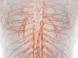
The lymphatic system is a network of vessels, nodes, and organs that helps maintain fluid balance and supports immune function. Lymphatic vessels collect excess fluid from tissues and eventually return it to the bloodstream.
In a lymphatic malformation, some of these vessels do not develop or connect normally. Lymph fluid becomes trapped inside abnormal channels and cyst-like spaces. The result may be a soft lump beneath the skin, clusters of tiny surface blisters, swelling in a limb, or a deeper mass that is invisible from the outside.
A lymphangioma is benign, meaning it is not cancer and does not spread through the body like a malignant tumor. That does not mean every case is harmless, however. A malformation in the tongue, throat, chest, abdomen, or near an eye can affect important structures even though its cells are noncancerous.
Why the terminology matters
Older records may use terms such as lymphangioma, cystic hygroma, cavernous lymphangioma, or lymphangioma circumscriptum. Modern vascular-anomaly specialists generally prefer “lymphatic malformation.” Using the current term can make it easier to find an experienced treatment center and avoid confusing the condition with hemangiomas, lymphomas, or other unrelated growths.
Types of Lymphatic Malformations
Lymphatic malformations are commonly described according to the size and pattern of their cystic spaces.
Macrocystic lymphatic malformations
Macrocystic LMs contain relatively large fluid-filled spaces. They may feel soft, compressible, or similar to a partially filled water balloon. Because the cysts are large enough to access with a needle, macrocystic lesions often respond well to image-guided sclerotherapy.
Microcystic lymphatic malformations
Microcystic LMs consist of many tiny abnormal channels that may spread between normal muscles, nerves, skin, and other tissues. On the skin or inside the mouth, they can appear as clusters of translucent or blood-tinged bumps. These lesions can be more difficult to treat because there is no single large cavity to drain or inject.
Mixed lymphatic malformations
A mixed LM contains both macrocystic and microcystic components. Treatment may combine several approaches because the large and small cysts do not always respond in the same way.
Specialists also distinguish isolated lymphatic malformations from complex lymphatic anomalies that affect multiple tissues, organs, bones, or central lymphatic channels. These uncommon disorders require particularly specialized evaluation.
Common Signs and Symptoms
Symptoms vary widely. A small superficial lesion may be mostly cosmetic, while a deeper malformation can produce significant functional problems.
Possible signs include:
- A soft or spongy lump under the skin
- Swelling that becomes larger during an infection
- Tiny clear, pink, purple, or blood-filled skin blisters
- Pain, warmth, redness, or tenderness
- Fluid leaking from surface vesicles
- Recurrent cellulitis or infection
- Bleeding into the malformation
- Limited movement when an arm, leg, or joint is involved
- Difficulty chewing, swallowing, speaking, or breathing
- Changes in vision when the tissues around an eye are affected
Lymphatic malformations may enlarge after viral illnesses, bacterial infections, physical trauma, internal bleeding, or hormonal changes. Families sometimes describe a lesion that seemed stable for months and then suddenly “inflated overnight.” The change does not automatically mean the condition has become cancerous, but sudden enlargement deserves medical attention.
How Is a Lymphangioma Diagnosed?
Medical history and physical examination
Diagnosis begins with a detailed history and examination. The clinician may ask when the swelling first appeared, whether it changes in size, whether infections have occurred, and whether the patient has pain, bleeding, breathing problems, or difficulty eating.
The malformation may be examined while the patient is sitting, lying down, swallowing, or moving the affected body part. Doctors also look for skin discoloration, warmth, tenderness, surface vesicles, asymmetry, and signs that other types of vessels are involved.
Ultrasound
Ultrasound is often the first imaging test for a superficial mass, especially in children. It does not use ionizing radiation, usually does not require sedation, and can show whether a lesion is fluid-filled or solid.
Doppler ultrasound can evaluate blood flow. A typical isolated lymphatic malformation has little or no internal flow, although small vessels may be present in its walls. This helps distinguish an LM from a high-flow arteriovenous malformation and from some vascular tumors.
Magnetic resonance imaging
MRI is usually the most useful test for mapping a large, deep, or complex lymphatic malformation. It can show how far the lesion extends and whether it surrounds muscles, nerves, blood vessels, the airway, or internal organs.
This information is essential before sclerotherapy or surgery. MRI may also reveal fluid levels caused by bleeding into the cysts. Younger children sometimes need sedation or anesthesia to remain still throughout the scan.
CT scans and other imaging
A CT scan may be used when rapid imaging is required, when bone or chest structures need evaluation, or when MRI is unavailable. Because CT uses radiation and generally provides less soft-tissue detail than MRI, it is not always the first choice for routine evaluation in children.
Specialized centers may use dynamic contrast MR lymphangiography or other advanced lymphatic imaging when abnormal lymphatic flow, chylous fluid leakage, or central conducting lymphatic disease is suspected.
Prenatal diagnosis
Some large lymphatic malformations are detected during pregnancy on prenatal ultrasound. Fetal MRI may then help define the lesion and determine whether it could obstruct the baby’s airway at delivery.
When a large neck malformation is found before birth, obstetricians, neonatologists, anesthesiologists, surgeons, and airway specialists may coordinate the delivery. Prenatal testing may also be offered because certain fetal neck malformations can occur with chromosomal or genetic conditions, although many lymphatic malformations occur on their own.
Biopsy and genetic testing
A biopsy is not routinely required when the clinical appearance and imaging are typical. It may be considered if the diagnosis is uncertain, the lesion behaves unusually, or doctors need to exclude another condition.
Genetic testing is becoming increasingly useful for extensive, recurrent, or syndromic malformations. Many lymphatic malformations are associated with genetic changes that occur only in the affected tissue rather than changes inherited from a parent. For this reason, testing a tissue or fluid sample may sometimes be more informative than testing blood alone.
Conditions That Can Resemble a Lymphangioma
A lump or patch of swelling should not be labeled a lymphangioma based on appearance alone. The differential diagnosis may include:
- Hemangioma or another vascular tumor
- Venous or venolymphatic malformation
- Branchial cleft cyst
- Thyroglossal duct cyst
- Dermoid or epidermoid cyst
- Abscess or infected lymph node
- Lipoma
- Hernia
- Benign or malignant soft-tissue tumor
The age of the patient, location of the mass, imaging characteristics, growth pattern, and blood-flow pattern help doctors separate these conditions. A rapidly growing or firm unexplained mass requires prompt evaluation rather than a confident diagnosis from a search-engine image gallery.
Does Every Lymphangioma Need Treatment?
No. Observation may be appropriate when a malformation is small, painless, stable, and not interfering with health or daily activities. Monitoring may include periodic examinations, photographs, measurements, and imaging when needed.
Treatment is more likely to be recommended when the lesion causes:
- Airway obstruction or breathing difficulty
- Problems with feeding, swallowing, speech, or vision
- Pain or repeated swelling
- Recurrent infection or cellulitis
- Bleeding or persistent fluid leakage
- Restricted mobility
- Compression of nerves or organs
- Significant disfigurement or emotional distress
The goal is not always to erase every abnormal channel. In complex cases, the more realistic goal may be controlling symptoms, protecting function, preventing complications, and reducing the malformation enough to improve quality of life.
Lymphangioma Treatment Options
Sclerotherapy
Sclerotherapy is a minimally invasive procedure commonly used for macrocystic and selected mixed lymphatic malformations. An interventional radiologist guides a needle into the cyst using ultrasound or other imaging. The fluid is removed, and a sclerosing medication is injected.
The medication irritates the abnormal lining of the cyst, causing inflammation and eventual scarring. As the treated space collapses, the malformation may shrink. The word “irritates” sounds alarming, but controlled irritation is the entire strategythe medical version of serving an eviction notice to a troublesome fluid pocket.
Swelling, soreness, bruising, and temporary enlargement are common after treatment. Several sessions may be needed, particularly when a lesion contains multiple separate cysts. The choice of sclerosing agent depends on the lesion, its location, the patient’s age, and the treatment center’s experience.
Surgery
Surgical excision may be considered when the malformation is localized and can be removed without unacceptable damage to nearby structures. Surgery may also be used when a lesion threatens the airway, causes major functional problems, repeatedly becomes infected, or does not respond adequately to less invasive treatment.
Complete removal is not always possible. Microcystic malformations can weave through nerves, muscles, salivary glands, and blood vessels like ivy through a fence. Attempting to remove every microscopic component may cause more harm than leaving a small amount behind.
Possible surgical risks include bleeding, infection, fluid collection, nerve injury, scarring, and recurrence. In some cases, staged debulking is safer than one extensive operation.
Laser and surface treatments
Laser treatment may help control superficial vesicles, bleeding, or lymphatic leakage on the skin or mucosal surfaces. Lasers do not usually eliminate a deep malformation, but they may improve troublesome surface symptoms.
Other local methods, including radiofrequency or electrosurgical techniques, may be used for selected microcystic lesions in specialized centers.
Medication
Systemic medication may be considered for extensive, infiltrative, recurrent, or life-threatening lymphatic malformations that cannot be adequately controlled with procedures alone.
Sirolimus, an mTOR-pathway inhibitor, has been used to reduce swelling, pain, bleeding, and functional problems in some complex vascular anomalies. It requires specialist supervision and laboratory monitoring because it can affect immune function, blood counts, cholesterol, wound healing, and other body systems.
Alpelisib targets the PI3K-alpha pathway. The FDA has approved alpelisib under the brand name Vijoice for adults and children age 2 and older who have severe manifestations of PIK3CA-related overgrowth spectrum and require systemic therapy. It is not a routine treatment for every small lymphatic malformation. Genetic findings, disease severity, potential benefits, and risks must all be considered.
Antibiotics and supportive care
Antibiotics treat bacterial infections involving the malformation or surrounding skin. They do not remove the LM itself. A suddenly red, hot, painful lesion accompanied by fever should be assessed promptly because cellulitis can progress quickly.
Depending on location and symptoms, supportive care may include compression garments, skin care, physical therapy, occupational therapy, speech therapy, dental care, nutrition support, pain management, or psychological counseling.
Recovery and Follow-Up
Recovery depends on the procedure and the lesion’s location. After sclerotherapy, swelling may temporarily make the area look worse before it looks better. This is especially important for head and neck lesions, where post-procedure swelling may affect the airway. Some patients need observation in the hospital.
After surgery, follow-up focuses on wound healing, nerve and muscle function, fluid accumulation, infection, and recurrence. Repeat imaging is not automatically necessary after every treatment; it is usually ordered when it will influence the next decision.
Lymphatic malformations can return or enlarge because untreated microscopic channels may remain. Recurrence does not necessarily mean that treatment failed. Many patients require a long-term management plan rather than one dramatic “before and after” procedure.
When to Seek Urgent Medical Care
Contact a medical professional promptly when a known or suspected lymphatic malformation suddenly changes. Emergency evaluation may be necessary for:
- Difficulty breathing, noisy breathing, or a feeling that the airway is narrowing
- Rapid swelling of the neck, tongue, face, or mouth
- Trouble swallowing saliva or fluids
- High fever with redness, warmth, or severe tenderness
- Rapid enlargement after bleeding or injury
- New weakness, numbness, vision changes, or severe pain
- Signs of dehydration in a child who cannot eat or drink
Head and neck lymphatic malformations deserve particular caution because even a benign lesion can become dangerous if swelling compresses the airway.
Choosing a Treatment Team
Complex lymphatic malformations are best evaluated by a multidisciplinary vascular-anomalies program. Depending on the location and severity, the team may include an interventional radiologist, pediatric surgeon, plastic surgeon, dermatologist, hematologist, geneticist, ear-nose-and-throat surgeon, orthopedic specialist, and rehabilitation therapist.
Useful questions to ask include:
- Is the diagnosis definitely a lymphatic malformation?
- Is it macrocystic, microcystic, mixed, or part of a complex anomaly?
- What structures does it involve?
- What happens if we observe it for now?
- What is the goal of treatment: cure, reduction, symptom control, or protection of function?
- How many procedures might be needed?
- Should tissue-based genetic testing be considered?
- What symptoms require urgent attention?
Conclusion
Lymphangioma, now more commonly called lymphatic malformation, is a noncancerous but sometimes complicated disorder of abnormal lymphatic vessels. Diagnosis usually combines a physical examination with ultrasound and MRI. Prenatal imaging, advanced lymphatic studies, biopsy, or genetic testing may be appropriate in selected cases.
Small, symptom-free lesions may simply be monitored. When treatment is necessary, options include sclerotherapy, surgery, laser therapy, antibiotics, supportive care, and targeted medication for certain severe or genetically defined conditions. The best plan is individualized because two malformations with the same name can behave very differently.
The central question is not, “How do we make the scan look perfect?” It is, “How do we protect breathing, movement, comfort, appearance, and everyday life with the least harmful treatment?” That question leads to better decisionsand usually fewer medical plot twists.
The Real-World Experience of Diagnosis and Treatment
For many patients and families, the first stage of the lymphangioma experience is uncertainty. A parent may notice that one side of a baby’s neck looks fuller, or an adult may discover a soft lump that seems to change size after a cold. Because lymphatic malformations are uncommon, the first explanation is not always the final one. The swelling may initially be called a cyst, birthmark, enlarged lymph node, infection, or hemangioma.
The diagnostic process often becomes clearer after referral to a vascular-anomalies center. Ultrasound may confirm that the mass contains fluid, while MRI shows its true boundaries. This can be a surprising moment. A bump that looks small from the outside may extend between several tissue layers. Conversely, a visually dramatic swelling may be composed of accessible cysts that respond well to treatment.
Waiting for appointments and imaging results can be emotionally difficult. The word “mass” tends to send the human imagination sprinting directly toward cancer. Learning that a lymphatic malformation is benign usually brings relief, followed quickly by a second question: “If it is benign, why does my child need surgery or anesthesia?” The answer is that location and function matter. A noncancerous lesion can still obstruct the airway, impair swallowing, limit movement, leak fluid, or become repeatedly infected.
Sclerotherapy is often described as minimally invasive, but the experience is not always minimal from the patient’s perspective. There may be fasting before anesthesia, several hours at the hospital, temporary swelling, soreness, and anxious inspection of the treated area afterward. Parents may worry when the lesion initially looks larger. Understanding in advance that inflammation is part of the treatment can make the recovery period less frightening.
Improvement may be gradual rather than cinematic. The lesion might shrink over several weeks, become softer, or cause fewer infections without disappearing completely. A second or third procedure may be recommended. This can feel discouraging until the treatment team explains that staged care is normal and often safer than an aggressive one-time intervention.
Surgery brings a different set of expectations. Families commonly hope the surgeon can “take the whole thing out.” Sometimes that is possible. In other cases, complete removal would risk damage to a facial nerve, major blood vessel, muscle, or salivary gland. Leaving a small portion behind may represent careful judgment rather than incomplete effort. Asking the surgeon what can be safely removedand what should intentionally remainhelps establish realistic expectations.
Daily life also matters. Children may face questions from classmates about visible swelling, scars, or compression garments. Adults may feel self-conscious during work meetings, photographs, dating, or exercise. Counseling, school communication, and peer support can be valuable parts of care, even though they do not appear on an MRI report.
Families often become excellent observers. They learn the lesion’s normal color and texture, what happens during a cold, and which changes signal infection or internal bleeding. Keeping photographs, symptom notes, medication lists, procedure reports, and imaging records can make future appointments more productive.
The long-term experience is usually less about pursuing perfection and more about building a reliable plan. That plan may include watching a stable lesion, treating infections quickly, scheduling procedures when symptoms justify them, and returning to normal activities whenever possible. With knowledgeable specialists and realistic expectations, many people with lymphatic malformations attend school, work, travel, play sports, raise families, and spend far more time living their lives than thinking about their diagnosis.
Medical note: This article is intended for general education and does not replace evaluation by a qualified healthcare professional. Treatment decisions should be made with a vascular-anomalies team familiar with the patient’s imaging, symptoms, medical history, and genetic findings. The content synthesizes guidance and research from major U.S. pediatric hospitals, NIH-indexed medical literature, rare-disease organizations, vascular-anomaly specialists, the ISSVA classification, and current FDA information.