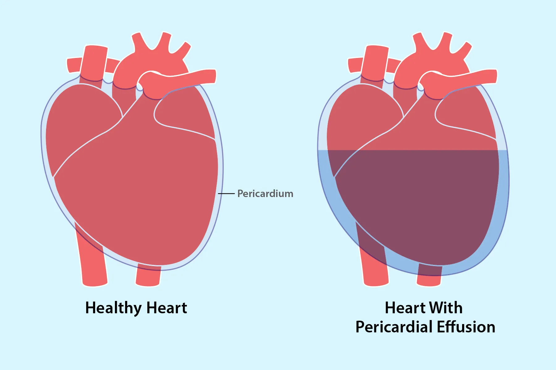
Pericardial effusion sounds like the kind of phrase a cardiologist says while everyone else quietly reaches for Google. In plain English, it means there is too much fluid in the sac around the heart. That sac is called the pericardium, and under normal circumstances, it contains a small amount of lubricating fluid that helps the heart move smoothly. Think of it as the heart’s nonstick pan coating. Helpful in small amounts. A problem when the kitchen floods.
Some pericardial effusions are small, quiet, and discovered by accident during an imaging test done for another reason. Others grow quickly or become large enough to squeeze the heart, making it harder for the heart chambers to fill and pump blood. That dangerous situation is called cardiac tamponade, and it is a medical emergency.
This guide explains the major pericardial effusion causes, common pericardial effusion symptoms, how doctors diagnose it, and what pericardial effusion treatment may involve. The goal is simple: clear information, no panic, and absolutely no pretending that “just drink more water” is a cardiology plan.
What Is Pericardial Effusion?
Pericardial effusion is the abnormal buildup of fluid in the pericardial space, the area between the heart and the pericardium. The pericardium is a thin, two-layered sac that surrounds the heart. Its job is to protect the heart, reduce friction, and help keep the heart in position inside the chest.
When extra fluid collects in this space, the effect depends on three big factors: how much fluid is present, how quickly it builds up, and whether the heart is being compressed. A slow-growing effusion may become fairly large before symptoms appear because the pericardium has time to stretch. A fast-growing effusion, even with less fluid, can cause serious pressure before the body has time to adapt.
Pericardial Effusion vs. Pericarditis
Pericarditis means inflammation of the pericardium. Pericardial effusion means fluid has accumulated around the heart. The two often travel together like an uninvited medical buddy system, but they are not identical. Pericarditis can cause an effusion, and an effusion can occur without obvious inflammation, depending on the underlying cause.
Common Causes of Pericardial Effusion
There is no single cause of fluid around the heart. Pericardial effusion can develop from infection, inflammation, injury, cancer, kidney disease, heart procedures, or certain medications. Sometimes, even after careful testing, the cause remains unknown. Doctors may call that an idiopathic pericardial effusion, which is medical-speak for “we looked hard, but the culprit left no business card.”
1. Infections
Viral infections are among the more common causes of pericardial inflammation and related effusion. Bacterial, fungal, and tuberculosis-related infections are less common in the United States but can be more severe. When infection is suspected, doctors may use blood tests, imaging, and sometimes fluid analysis to identify the source.
2. Inflammatory and Autoimmune Conditions
Autoimmune diseases can cause the immune system to attack or inflame tissues around the heart. Conditions such as lupus, rheumatoid arthritis, and other systemic inflammatory diseases may contribute to pericarditis and pericardial effusion. Treatment usually focuses on controlling inflammation and managing the underlying disease.
3. Cancer
Some cancers can spread to the pericardium or trigger fluid buildup around the heart. Lung cancer, breast cancer, lymphoma, and other malignancies may be associated with pericardial effusion. A malignant pericardial effusion often requires close monitoring and may need drainage, lab testing of the fluid, and cancer-directed therapy.
4. Kidney Failure
Advanced kidney disease can lead to uremic pericarditis, where waste products build up in the blood and irritate the pericardium. In these cases, improving kidney-related treatment, such as dialysis management, may be part of the overall plan.
5. Heart Attack, Heart Surgery, or Medical Procedures
Pericardial effusion may occur after a heart attack, cardiac surgery, catheter-based procedures, pacemaker placement, or chest trauma. Post-procedure effusions may be small and monitored, but rapidly accumulating blood or fluid around the heart requires urgent evaluation.
6. Hypothyroidism and Other Medical Conditions
Severe underactive thyroid disease can sometimes cause pericardial effusion. Other possible contributors include radiation therapy to the chest, certain medications, and systemic illnesses. Because the list is long, the medical history mattersa lot.
Pericardial Effusion Symptoms
Small pericardial effusions may cause no symptoms at all. A person might feel completely normal, then learn about the effusion after an echocardiogram, CT scan, or chest imaging test. Larger or faster-growing effusions are more likely to cause noticeable symptoms.
Common Symptoms
Possible symptoms of pericardial effusion include:
- Chest pain, pressure, or discomfort
- Shortness of breath, especially when lying flat
- Fast heartbeat or heart palpitations
- Fatigue or unusual weakness
- Lightheadedness, dizziness, or fainting
- Dry cough
- Swelling in the legs or abdomen in some cases
- A feeling of fullness or pressure in the chest
Chest pain from pericardial inflammation may feel sharp and may worsen when breathing deeply, coughing, or lying down. Some people feel better when sitting up and leaning forward. However, chest symptoms can come from many causes, including heart attack, lung problems, acid reflux, and muscle strain. Translation: the chest is not a great place for guessing games.
Emergency Warning Signs
Seek emergency medical care right away if symptoms include severe chest pain, trouble breathing, fainting, bluish or gray skin color, confusion, extreme weakness, or a very fast heartbeat at rest. These may be signs of cardiac tamponade or another serious condition.
What Is Cardiac Tamponade?
Cardiac tamponade happens when pressure from fluid around the heart prevents the heart chambers from filling properly. If the heart cannot fill, it cannot pump enough blood to the body. This can cause low blood pressure, shock, and life-threatening organ damage.
Doctors may look for signs such as low blood pressure, rapid heartbeat, swollen neck veins, muffled heart sounds, and pulsus paradoxus, which is an abnormal drop in blood pressure during inhalation. Not every person has every classic sign, so clinicians rely on the full picture: symptoms, physical exam, imaging, and hemodynamic stability.
Cardiac tamponade is not treated with “wait and see.” It usually requires urgent drainage of the fluid, often with a procedure called pericardiocentesis or, in certain cases, surgery.
How Doctors Diagnose Pericardial Effusion
Diagnosis starts with a conversation and a physical exam. A healthcare professional will ask about symptoms, recent infections, autoimmune disease, cancer history, kidney disease, heart procedures, medications, and chest injury. Then comes the detective work, but with more stethoscopes and fewer trench coats.
Physical Exam
During the exam, the clinician may listen for abnormal heart sounds or a pericardial friction rub, which can occur when inflamed pericardial layers rub against each other. They may also check blood pressure, pulse, oxygen level, neck veins, and signs of fluid retention.
Echocardiogram
An echocardiogram, or heart ultrasound, is the key test for confirming pericardial effusion. It can show the presence of fluid, estimate the size of the effusion, and help determine whether the heart is being compressed. If tamponade is suspected, echocardiography is often performed urgently.
Electrocardiogram
An electrocardiogram, also called an ECG or EKG, records the heart’s electrical activity. In pericardial effusion, it may show low-voltage QRS complexes or, in large effusions, electrical alternans, where the electrical pattern appears to vary as the heart swings slightly in the fluid-filled sac.
Chest X-Ray
A chest X-ray may show an enlarged heart silhouette when the effusion is large. However, a chest X-ray cannot reliably confirm small effusions or measure pressure on the heart. It is useful, but it is not the whole story.
CT Scan and Cardiac MRI
CT scans and cardiac MRI can provide additional detail, especially when the effusion is loculated, when the pericardium appears thickened, or when doctors need a better view of surrounding structures. MRI may also help evaluate pericardial inflammation and constrictive pericarditis.
Blood Tests and Fluid Analysis
Blood tests may check inflammation markers, kidney function, thyroid function, infection signs, autoimmune markers, or heart muscle injury. If fluid is drained, it may be sent to a lab for analysis. Fluid testing can help identify infection, cancer cells, bleeding, or other clues about the cause.
Pericardial Effusion Treatment
Treatment depends on the cause, size, symptoms, and whether the effusion is affecting heart function. A small, stable effusion may only need monitoring and treatment of the underlying condition. A large or symptomatic effusion may require medication, drainage, or surgery.
Monitoring
If the effusion is small and the patient is stable, doctors may recommend follow-up visits and repeat echocardiograms. Monitoring helps make sure the fluid is not increasing or causing pressure on the heart.
Medications
When inflammation is the main driver, treatment may include nonsteroidal anti-inflammatory drugs, colchicine, or sometimes corticosteroids. If a bacterial infection is present, antibiotics may be needed. If hypothyroidism, kidney disease, or autoimmune disease is responsible, treating that condition becomes central to the plan.
Pericardiocentesis
Pericardiocentesis is a procedure in which a doctor drains fluid from the pericardial space using a needle and catheter, usually guided by imaging. It may be performed urgently for cardiac tamponade or diagnostically when the cause of the effusion is unclear. The drained fluid may be tested in a lab.
Pericardial Window or Surgery
If fluid keeps coming back, is difficult to drain, or is related to certain cancers or infections, doctors may recommend a pericardial window. This surgical procedure creates an opening that allows fluid to drain from the pericardial space into another area of the chest or abdomen where the body can absorb it more easily.
Treating the Underlying Cause
The most effective treatment is not just removing fluid; it is solving why the fluid appeared in the first place. For example, a viral inflammatory effusion may improve with anti-inflammatory therapy and rest, while a cancer-related effusion may require oncology care. A kidney-related effusion may improve with better dialysis management. One-size-fits-all medicine belongs in the same drawer as one-size-fits-all jeans: suspicious.
Recovery and Outlook
The outlook for pericardial effusion varies widely. Many small or inflammation-related effusions improve with treatment and monitoring. Effusions caused by bacterial infection, cancer, trauma, or severe systemic disease may be more serious and require more aggressive care.
Recovery may include follow-up echocardiograms, medication adjustments, activity guidance, and monitoring for recurrence. Some people recover quickly; others need ongoing care from cardiology, oncology, rheumatology, nephrology, or another specialist depending on the cause.
Can Pericardial Effusion Be Prevented?
Not every case can be prevented. However, good medical management of chronic conditions can reduce risk. That includes treating infections promptly, managing autoimmune disease, following kidney disease care plans, taking thyroid disease seriously, and attending follow-up appointments after heart surgery or cardiac procedures.
Prevention also means listening to symptoms. New chest pain, unexplained shortness of breath, fainting, or a racing heartbeat should not be treated like a random software glitch. The human body does not come with a “restart and ignore” button.
Living With or Recovering From Pericardial Effusion: Practical Experiences
People who go through pericardial effusion often describe the experience as confusing because symptoms can be vague. One person may feel sharp chest pain and shortness of breath. Another may simply feel tired, winded on stairs, or uncomfortable when lying flat. A third may have no symptoms and discover the condition during imaging for something else entirely. That variety can make pericardial effusion feel mysterious, but it is exactly why medical evaluation matters.
A common experience is the “I thought I was just out of shape” moment. Someone may notice that walking across a parking lot suddenly feels harder than usual. They may blame stress, poor sleep, or too much coffee. Sometimes those are reasonable suspects. But when shortness of breath is new, worsening, or paired with chest pressure, dizziness, or a fast heartbeat, it deserves attention.
Another experience involves anxiety after diagnosis. Hearing that there is fluid around the heart can sound terrifying. Many patients immediately imagine the worst. In reality, the seriousness depends on the size of the effusion, how quickly it formed, the cause, and whether it is affecting heart function. A small, stable effusion may be monitored. A large effusion or tamponade requires urgent treatment. The same diagnosis can lead to very different care plans, which is why follow-up questions are useful.
Patients often learn new vocabulary quickly: echocardiogram, pericardium, tamponade, inflammatory markers, pericardiocentesis. It can feel like being dropped into a medical spelling bee with no warning. A helpful strategy is to ask the care team three simple questions: How large is the effusion? Is it affecting how my heart pumps? What do we think caused it? These questions turn a scary label into a clearer action plan.
After treatment, many people are advised to watch for returning symptoms. This may include chest pain, worsening shortness of breath, fainting, swelling, fever, or a racing heartbeat. Follow-up imaging may be scheduled to confirm that the fluid is stable or improving. For inflammation-related cases, taking medications exactly as prescribed matters because stopping early may increase the chance of symptoms returning.
Daily life after pericardial effusion may involve temporary activity limits. Some people need rest while inflammation settles. Athletes and highly active individuals may need specific clearance before returning to intense exercise. That can be frustrating, especially for people who use workouts to manage stress. But the heart is one muscle you do not want to negotiate with like a stubborn gym coach.
Emotionally, the experience can also change how people relate to routine symptoms. A skipped heartbeat or mild chest twinge may suddenly feel alarming. That reaction is understandable. The goal is not to panic at every sensation, but to understand which symptoms require urgent care and which can be discussed at follow-up. A written plan from the healthcare team can reduce uncertainty.
Family members and caregivers can help by listening, driving to appointments, taking notes, and encouraging follow-up care. They do not need to become amateur cardiologists. In fact, please do not let the family group chat become a cardiology board review. The best support is practical: help track medications, symptoms, appointments, and questions for the doctor.
The biggest takeaway from real-life experiences is this: pericardial effusion is not a single story. It can be mild or serious, temporary or recurring, obvious or silent. The safest path is timely evaluation, cause-based treatment, and consistent follow-up. With the right care plan, many people recover well and return to normal routines with a better understanding of their heart health.
Conclusion
Pericardial effusion is a buildup of extra fluid around the heart. Sometimes it is small and harmless enough to monitor. Sometimes it is a warning sign of infection, inflammation, cancer, kidney disease, thyroid disease, trauma, or complications after heart procedures. The key is not simply knowing that fluid exists; it is knowing why it is there and whether it is affecting the heart’s ability to pump.
Symptoms such as chest pain, shortness of breath, dizziness, fainting, and a fast heartbeat should be taken seriously, especially if they appear suddenly or worsen. Echocardiography is the central diagnostic test, while blood tests, ECG, chest X-ray, CT, MRI, and fluid analysis may help identify the cause. Treatment may involve monitoring, anti-inflammatory medicine, antibiotics, management of underlying disease, pericardiocentesis, or surgery.
If there is one practical message to remember, it is this: fluid around the heart is not a DIY project. It deserves professional evaluation, especially when symptoms suggest cardiac tamponade. The heart works hard enough already; it should not have to perform under pressure from an unwanted fluid roommate.