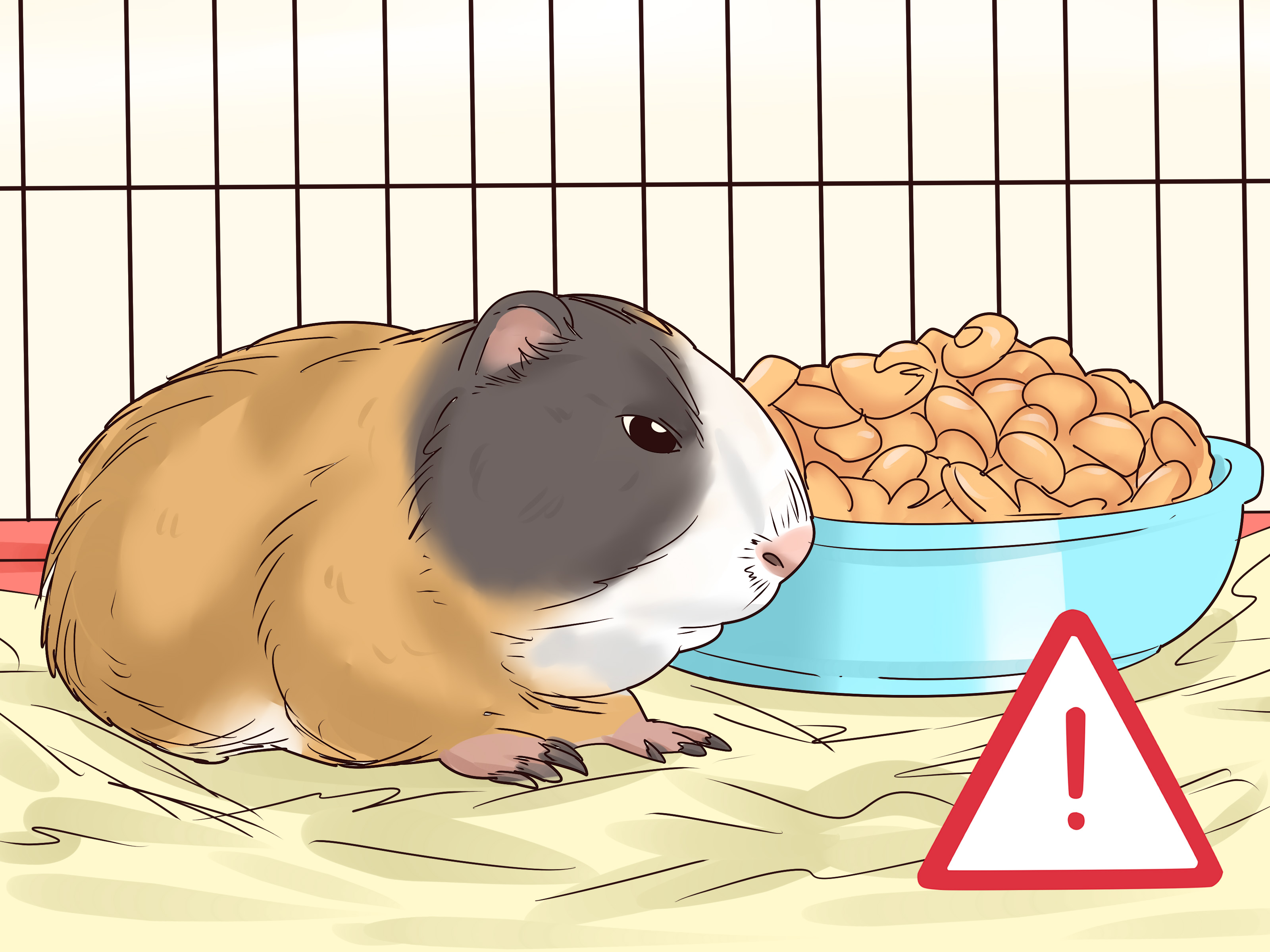
Guinea pigs are adorable, opinionated little hay machines. They squeak for snacks, redecorate their bedding like tiny interior designers, and somehow manage to look offended when you are two minutes late with lettuce. But when you find a lump on one of these furry potatoes, the mood changes fast. Suddenly, every bump feels suspicious, every nap looks dramatic, and every internet search turns into a stress marathon.
Here is the good news: not every lump in a guinea pig is cancer, and not every tumor is a disaster. Some masses are benign. Some can be removed successfully. Some are not even tumors at all, but abscesses, swollen lymph nodes, cysts, or reproductive problems pretending to be something else. The trick is to stop guessing, start evaluating, and let a guinea pig-savvy veterinarian sort out what is actually going on.
This guide breaks the process into three practical ways to diagnose and treat tumors in guinea pigs. Think of it as a roadmap: first notice the problem, then confirm what it is, then choose the best treatment for both the mass and the pig attached to it. Because in exotic pet medicine, the lump matters, but the whole patient matters more.
Why Guinea Pig Tumors Can Be So Tricky
Tumors in guinea pigs tend to show up more often as they age, especially after about three years old. Common sites include the skin, mammary glands, lungs, ovaries, uterus, and the blood or lymphatic system. Some skin masses are benign and slow-growing. Mammary masses can be benign in some cases and malignant in others. Reproductive tumors may hide inside the abdomen until the belly becomes enlarged. Lung tumors can look suspiciously like a respiratory infection. In other words, guinea pig medicine loves a plot twist.
That is exactly why home diagnosis is a bad plan. A firm bump on the rump could be a benign skin tumor. A swelling under the jaw could be an abscess or an enlarged lymph node. A distended belly could point toward an ovarian or uterine problem, but it could also mean something else entirely. Looking at a lump and declaring, “Yep, definitely cancer,” is a little like diagnosing a car problem by tapping the hood and nodding wisely. It feels confident, but it is not very useful.
Way 1: Start With the Lump You Can See and the Symptoms You Cannot
Pay attention to size, location, and behavior
The first step is simple but important: notice what changed. Many owners first detect a tumor by feeling a firm nodule while petting their guinea pig, spotting swelling around a mammary gland, or noticing a lump near the rump or lower back. Skin tumors in guinea pigs are often round and firm. Mammary tumors may show up as swelling in one or more glands and can sometimes leak clear or bloody fluid. Internal tumors are sneakier, but they can cause weight loss, a swollen abdomen, reduced appetite, breathing trouble, lethargy, or changes in grooming.
At home, your job is not to diagnose the mass. Your job is to document it like a very worried but organized assistant manager. Take a photo. Note the date you found it. Measure it gently if you can. Watch whether it is growing, ulcerating, bleeding, or interfering with movement, eating, or bathroom habits. That information can help your veterinarian decide how urgent the situation is.
Know the red flags that mean “book the vet now”
Some changes deserve same-day or urgent attention. These include rapid growth, open sores, discharge, trouble breathing, refusal to eat, sudden weight loss, pain when touched, bloody vaginal discharge, or a belly that seems to enlarge over days rather than weeks. Guinea pigs are prey animals, which means they are talented at pretending everything is fine until everything is absolutely not fine. By the time a guinea pig looks obviously sick, the problem may be more advanced than it appears.
Also remember that some non-tumor problems can look almost identical to cancer at first glance. Abscesses are a classic example. Guinea pigs form thick pus that often does not drain well, so an abscess can feel like a solid mass. Enlarged lymph nodes can also confuse the picture. That is one more reason every suspicious lump deserves a professional exam instead of a household debate featuring zero veterinarians and one panicked search history.
Get a hands-on exam from an exotics veterinarian
The best starting point is a veterinarian with experience in exotics or small mammals. These vets are more likely to recognize guinea pig-specific tumor patterns, anesthesia concerns, and medication issues. During the exam, the vet will assess the lump’s mobility, consistency, depth, and relationship to nearby tissues. They will also check body condition, breathing, heart rate, lymph nodes, abdomen, mammary tissue, mouth, and skin. A tiny mass can be the tip of a much larger medical iceberg, so the full-body exam matters.
Way 2: Confirm the Diagnosis With Tests Instead of Guesswork
Use fine-needle aspiration, biopsy, and pathology
Once a suspicious mass is found, the next step is figuring out what it actually is. Fine-needle aspiration is often one of the first tools. This uses a thin needle to collect cells from the lump so they can be examined under a microscope. It can help distinguish inflammation from neoplasia and may provide a quick clue about whether the lesion looks benign, malignant, or infected.
But cytology has limits. Some tumors do not shed many cells, and some samples are messy, bloody, or frustratingly unhelpful. That is why biopsy and histopathology are often the gold standard. A biopsy can involve removing part of the mass or taking the whole lump out and sending it to a pathologist. Histopathology looks at tissue architecture, not just loose cells, which gives a more reliable diagnosis and helps answer the questions owners actually care about: What is it? Is it benign or malignant? Are the margins clean? Could it spread?
This is especially important for mammary masses. Veterinary sources do not all agree on how often guinea pig mammary tumors are benign versus malignant, which tells you something important: appearance alone is not enough. A squishy lump is not automatically harmless, and a firmer one is not automatically cancer. Pathology is the referee here, and yes, the referee should get the final whistle.
Stage the tumor with imaging and lab work
If the lump is internal, large, fast-growing, or suspicious for malignancy, your veterinarian may recommend additional tests. These can include bloodwork, radiographs, ultrasound, and sometimes CT. Imaging helps answer big questions: Is the mass attached to deeper tissue? Has it spread to the lungs? Are lymph nodes enlarged? Is the belly enlargement coming from the uterus, ovaries, or another organ? Is that “neck lump” a thyroid mass, an abscess, or an enlarged node?
Thoracic radiographs are often used to look for spread to the lungs. Ultrasound can help assess abdominal masses and guide aspirates. CT may be recommended when the anatomy is complicated or when surgeons need a better map before operating. Bloodwork does not diagnose every tumor directly, but it helps assess organ function, inflammation, anemia, hydration, and whether a guinea pig is a safer candidate for anesthesia and surgery.
Do not skip the differential diagnosis
A good vet does not just ask, “What tumor is this?” They ask, “What else could this be?” In guinea pigs, that list may include abscesses, cysts, ovarian disease, uterine disease, enlarged lymph nodes, leukemia, and infectious conditions. This is not overthinking. It is good medicine. A surgery plan based on the wrong assumption can waste time, money, and recovery energy on the wrong problem.
Way 3: Treat the Tumor and the Guinea Pig, Not Just the Lump
Surgery is often the main event
For many skin and mammary tumors, surgery is the primary treatment. If the mass is localized and removable, surgical excision may be curative. In skin tumors, this can be straightforward when the lump is small and well-defined. In mammary tumors, wider excision may be recommended, sometimes along with nearby lymph node evaluation. If the tumor is ulcerated, painful, or interfering with movement, removing it can improve comfort even when cure is uncertain.
That said, guinea pigs are not tiny dogs in fuzzy pants. They have specific anesthetic and surgical needs. They can become cold quickly, they do not handle stress especially well, and they need careful fluid support, pain control, and close monitoring before, during, and after surgery. An exotics-focused team is a major advantage here because details such as warming, feeding support, species-safe antibiotics, and gastrointestinal monitoring can make a huge difference in recovery.
When advanced cancer care enters the conversation
If the tumor is malignant or has spread, treatment may go beyond surgery. Depending on the case, a veterinarian may discuss chemotherapy, radiation therapy, or referral to an exotic animal specialty hospital. These options are not appropriate or available for every guinea pig, but they do exist. In some published veterinary cases, advanced diagnostics and specialty care have helped confirm unusual tumors such as thyroid neoplasia or metastatic disease.
Sometimes, however, the most humane plan is not the fanciest one. If a tumor is widespread, nonresectable, or causing major systemic illness, palliative care may be the best route. That means focusing on comfort, appetite, pain control, hydration, clean housing, and quality of life. There is nothing lazy or lesser about that choice. Sometimes good medicine means fighting hard, and sometimes it means choosing kindness with a very clear head.
Supportive care matters more than many owners expect
Guinea pigs recovering from tumor treatment often need nursing support. That can include pain medication, syringe feeding if appetite drops, careful weight checks, soft bedding, wound monitoring, and follow-up visits. A guinea pig that stops eating after surgery is not “just being moody.” Reduced appetite can spiral into gastrointestinal trouble quickly, so early intervention is important.
Owners should also monitor for recurrence. Some tumors can regrow if they were not fully removed, and malignant tumors may metastasize even after a successful-looking surgery. Recheck appointments are not optional fluff. They are part of the treatment plan.
Common Tumor Patterns Owners Should Know
While only a veterinarian can diagnose a specific case, a few tumor patterns show up often enough to be worth recognizing.
- Skin tumors: These are among the most common external masses in guinea pigs. Many are benign, including some hair follicle tumors, but they still need evaluation and often surgical removal.
- Mammary tumors: These can occur in both males and females. Some are benign fibroadenomas, while others are malignant adenocarcinomas or fibroadenocarcinomas. Pathology is essential.
- Reproductive tumors: Uterine and ovarian tumors may cause abdominal enlargement, discomfort, or bloody discharge. Early spaying can reduce some reproductive tumor risks in females.
- Lymphoid tumors: Leukemia and lymphoma may cause enlarged nodes, rough coat, lethargy, weight loss, and systemic illness. These can progress quickly.
- Lung tumors: Some guinea pigs develop lung tumors that mimic pneumonia, causing coughing, sneezing, lethargy, and breathing difficulty.
- Thyroid tumors: Less common but increasingly recognized, these may be linked with weight loss and a cervical mass and may require advanced imaging or specialty care.
How to Lower Risk and Catch Problems Earlier
You cannot guarantee your guinea pig will never develop a tumor, but you can improve the odds of catching trouble early. Monthly at-home wellness checks help you notice new lumps before they become giant unwelcome roommates. Regular weigh-ins matter because unexplained weight loss is often the first clue that something internal is wrong. Senior guinea pigs benefit from consistent veterinary well visits, and females not intended for breeding may benefit from discussing early spaying with an exotics veterinarian because it can reduce the risk of some reproductive diseases and tumors.
Good husbandry helps too. Clean housing, high-quality hay, a balanced guinea pig diet, and routine observation will not magically repel every tumor in the universe, but they do make it easier to spot abnormal changes sooner. And sooner is usually better in oncology, whether your patient weighs sixty pounds or sixty ounces.
Experiences Owners Commonly Have With Guinea Pig Tumors
In real life, the experience of dealing with a guinea pig tumor rarely begins with a dramatic movie moment. It usually starts with something small and weird. An owner is doing a routine cuddle check and notices a pea-sized bump near the rump. Or they are trimming nails and feel a soft swelling near a mammary gland. Or their usually enthusiastic lettuce enthusiast begins eating more slowly, losing weight, or making breathing noises that sound like a squeaky toy with an attitude problem.
One of the most common experiences owners describe is confusion. The guinea pig still begs for snacks. He still argues with the vacuum cleaner. She still rearranges hay like it is part of a personal design philosophy. Because the pet is still acting partly normal, it is easy to hope the lump is “probably nothing.” Then the bump grows. Or it becomes irritated. Or a vet visit reveals that the external lump was only one piece of a larger problem. The lesson many owners learn is simple: in guinea pigs, small changes deserve attention because these animals are masters of underreporting their own health problems.
Another common experience is surprise at how much diagnostics matter. Many owners go into the appointment expecting the vet to identify the lump on sight. Instead, they hear words like aspirate, biopsy, radiographs, ultrasound, histopathology, margins, and staging. It can feel overwhelming at first. But owners who go through the process often realize that testing replaces panic with information. Instead of living in the land of “maybe,” they get an actual diagnosis and a real treatment plan. That alone can be a huge emotional relief.
Owners also frequently underestimate recovery. A guinea pig who has surgery may need more hands-on support than expected. There may be syringe feeding, medication schedules, bedding changes, and daily incision checks. Some guinea pigs bounce back quickly and act personally offended that they were ever taken to the hospital. Others need more encouragement, more calories, and more careful monitoring. People often come away from the experience with a new respect for how delicate, but also how resilient, these little animals can be.
Then there is the emotional side. Some owners get the best possible news: the lump is benign, the surgery margins are clean, and their guinea pig returns to normal life with only a scar and an even stronger opinion about vegetables. Others get harder news, especially with lymphoma, internal tumors, or metastatic disease. In those cases, owners often say the most meaningful part of the experience was having a clear explanation and a plan that focused on comfort and quality of life. Even when the ending is not the one anyone wanted, good veterinary guidance helps families make thoughtful, loving decisions instead of reactive ones.
The biggest shared lesson is probably this: guinea pig tumors are scary, but they are not a reason to give up at first sight. Some masses are treatable. Some are curable. Some can be managed. And even when cure is not possible, comfort still is. The owners who handle these situations best are usually not the ones with the fanciest internet theories. They are the ones who noticed changes early, sought real veterinary help, asked smart questions, and kept showing up for their tiny patient with hay, patience, and a lot of heart.
Conclusion
If you remember only one thing from this article, make it this: a lump in a guinea pig is never a “wait and see forever” kind of problem. The three best ways to diagnose and treat tumors in guinea pigs are to catch changes early, confirm the diagnosis with proper veterinary testing, and choose treatment based on the type of tumor and the condition of the whole animal. That might mean surgery. It might mean advanced imaging or oncology care. It might mean palliative support. But it should always begin with a real exam and a real plan.
Guinea pigs may be small, but their medical problems are not. A timely diagnosis can turn a frightening bump into a manageable problem, and in some cases, a curable one. So check the fluff, trust the diagnostics, and never underestimate what a determined little cavy and a good exotics vet can do together.