Cardiac tamponade is a life-threatening emergency in which fluid or blood builds up inside the sac surrounding the heart. As pressure rises, the heart cannot fill normally between beats. Less blood is pumped to the body, blood pressure may fall, and vital organs can quickly become deprived of oxygen.
In plain English, the heart is being squeezed inside a space that cannot expand fast enough. The heart is remarkably hardworking, but it does not perform well when it is being shrink-wrapped.
Cardiac tamponade can develop suddenly after an injury or medical procedure, or more gradually because of inflammation, cancer, kidney disease, or another medical condition. Either way, suspected tamponade requires immediate hospital treatment. Anyone experiencing severe shortness of breath, fainting, sudden chest discomfort, confusion, or signs of shock should call 911 rather than attempting to diagnose the problem at home.
What is cardiac tamponade?
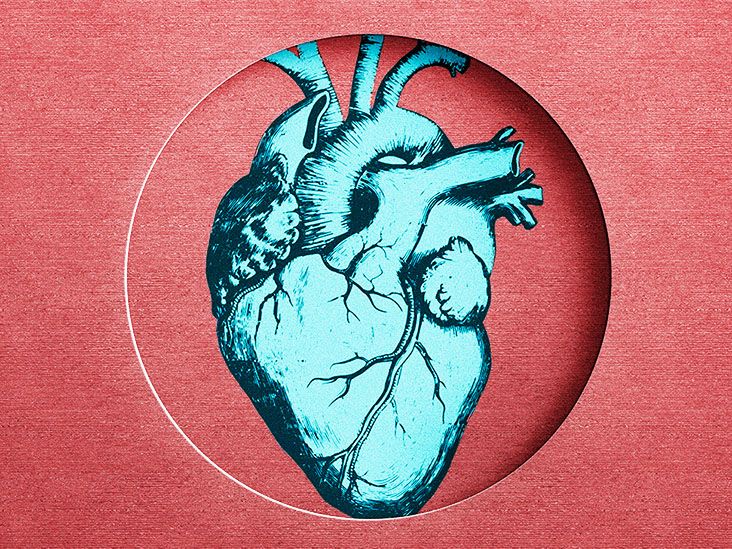
The heart sits inside a protective, double-layered sac called the pericardium. A small amount of lubricating fluid normally lies between these layers, allowing the heart to beat without creating excessive friction.
A pericardial effusion occurs when additional fluid collects in that space. Not every effusion causes cardiac tamponade. Tamponade develops when pressure from the accumulating fluid becomes high enough to interfere with the heart’s ability to fill and pump.
Why the speed of fluid buildup matters
The seriousness of an effusion is not determined by volume alone. When blood or fluid accumulates rapidly, even a relatively modest amount can create dangerous pressure because the pericardium has little time to stretch. This can happen after penetrating chest trauma, heart surgery, a catheter-based procedure, or a tear involving the heart or aorta.
When fluid accumulates gradually over weeks or months, the pericardium may stretch and temporarily accommodate a much larger volume. Symptoms may therefore appear slowly. Once the sac reaches its limit, however, the patient’s condition can deteriorate quickly.
The pressure first affects the thinner-walled right side of the heart. The right atrium and right ventricle cannot fill normally, so less blood reaches the lungs and subsequently the left side of the heart. Cardiac output falls, producing a form of circulatory failure called obstructive shock.
What causes cardiac tamponade?
Anything that causes blood, inflammatory fluid, pus, or other material to collect around the heart can potentially lead to tamponade. Some causes produce a sudden emergency, while others create a slower buildup.
Chest injuries
Blunt trauma from a vehicle collision, fall, or forceful impact can damage the heart or nearby blood vessels. Penetrating injuries, including stab or gunshot wounds, may cause blood to rapidly enter the pericardial sac. Traumatic tamponade can progress within minutes and may require emergency surgery.
Complications of medical procedures
Cardiac tamponade is an uncommon but recognized complication of procedures involving the heart and major blood vessels. Examples include:
- Cardiac catheterization or coronary intervention
- Pacemaker or defibrillator lead placement
- Electrophysiology studies and catheter ablation
- Heart biopsy
- Central venous catheter placement
- Open-heart surgery
These procedures are generally performed safely, but an accidental perforation can allow blood to collect around the heart. Hospital teams monitor patients so that this complication can be identified and treated promptly.
Pericarditis and infections
Pericarditis is inflammation of the pericardium. It may follow a viral illness, bacterial infection, tuberculosis, heart attack, surgery, or an immune-system reaction. Inflammation can trigger a pericardial effusion, which occasionally progresses to tamponade.
Purulent pericarditis, in which bacteria cause pus to collect in the pericardial space, is particularly serious. It requires drainage plus targeted antimicrobial treatment.
Cancer
Cancers of the lung, breast, blood, lymphatic system, and other organs can spread to the pericardium or interfere with normal fluid drainage. Malignant effusions may return after initial drainage, so some patients need a surgical pericardial window or another procedure designed to reduce recurrence.
Kidney failure
Advanced kidney disease can cause uremic pericarditis and fluid accumulation around the heart. Treatment may involve urgent drainage when tamponade is present, along with dialysis and management of the underlying kidney disorder.
Autoimmune and inflammatory diseases
Lupus, rheumatoid arthritis, and other inflammatory conditions can affect the pericardium. Controlling the underlying disease helps reduce the likelihood of continued inflammation and recurrent fluid buildup.
Other possible causes
- Aortic dissection
- Rupture of the heart wall after a heart attack
- Severe hypothyroidism
- Radiation therapy involving the chest
- Bleeding associated with anticoagulant medication
- Rare reactions to medicines
- An unexplained or idiopathic pericardial effusion
Anticoagulants do not automatically cause tamponade, but they can make bleeding more severe when an injury or perforation occurs. Patients should never stop prescribed blood thinners without medical guidance.
Cardiac tamponade symptoms
Symptoms vary according to how quickly pressure develops and how much the circulation has been affected. Rapid tamponade may cause dramatic collapse. Slowly developing tamponade can resemble heart failure, lung disease, or severe fatigue.
Common warning signs
- Shortness of breath or unusually rapid breathing
- Chest pressure, fullness, or pain
- A fast or pounding heartbeat
- Dizziness or lightheadedness
- Fainting or nearly fainting
- Extreme weakness
- Anxiety, restlessness, or a sense that something is seriously wrong
- Difficulty breathing while lying flat
- Swelling of the abdomen, legs, or neck veins
- Reduced urination
- Pale, cool, gray, or bluish skin
- Confusion or reduced alertness
Chest pain may spread to the shoulder, back, arm, or neck. If pericarditis is involved, the pain may worsen with a deep breath or while lying flat and improve when the person sits up and leans forward. These patterns are helpful clues, but they cannot safely distinguish tamponade from a heart attack, pulmonary embolism, aortic dissection, or another emergency.
Beck’s triad
Clinicians traditionally associate cardiac tamponade with three findings known as Beck’s triad:
- Low blood pressure
- Distended neck veins
- Muffled or distant heart sounds
The triad is memorable, but it is not present in every patient. Waiting for all three findings would be like waiting for every warning light on a dashboard before admitting the engine has a problem.
Pulsus paradoxus and other examination findings
A clinician may detect pulsus paradoxus, an abnormally large drop in systolic blood pressure during inhalation. Other possible findings include a rapid pulse, narrow pulse pressure, cool extremities, weak peripheral pulses, and signs of poor organ perfusion.
People with slowly developing tamponade may initially maintain a normal blood pressure because the body compensates by increasing the heart rate and tightening blood vessels. A normal reading therefore does not completely rule out an evolving problem.
When cardiac tamponade is an emergency
Cardiac tamponade is always potentially serious. Call 911 immediately when chest discomfort or breathing difficulty occurs with fainting, confusion, heavy sweating, blue or gray skin, a very rapid heartbeat, or severe weakness.
Emergency evaluation is especially important after chest trauma, heart surgery, catheter ablation, pacemaker placement, cardiac catheterization, or another invasive procedure. Do not drive yourself when symptoms are severe. An ambulance team can begin monitoring and emergency support while traveling to the hospital.
How cardiac tamponade is diagnosed
Diagnosis combines the patient’s medical history, physical examination, vital signs, and imaging. Treatment may begin before every test is completed when the patient is unstable.
Echocardiography
An echocardiogram uses ultrasound to show the heart, surrounding fluid, chamber movement, and changes in blood flow. It is the central imaging test for suspected tamponade because it can be performed rapidly at the bedside.
Clinicians look for a pericardial effusion, collapse of the right atrium or right ventricle during parts of the heartbeat, changes in blood flow with breathing, and a swollen inferior vena cava that does not collapse normally. Ultrasound can also help guide a drainage needle.
Electrocardiogram
An electrocardiogram, or ECG, records the heart’s electrical activity. It may show a fast heart rate, low-voltage signals, or electrical alternans, a pattern in which the electrical signal varies as the heart moves within a large effusion. These findings can support the diagnosis but are not present in every case.
Chest X-ray and advanced imaging
A chest X-ray may show an enlarged, rounded cardiac silhouette when a large effusion has accumulated gradually. It may appear normal when blood collects rapidly, so a normal X-ray does not exclude acute tamponade.
Computed tomography or cardiac magnetic resonance imaging can provide detailed information about the pericardium and nearby structures in stable patients. Advanced imaging should not delay emergency drainage when shock is developing.
Blood and fluid testing
Laboratory tests may evaluate anemia, infection, kidney function, thyroid function, inflammation, heart-muscle injury, and clotting. After drainage, pericardial fluid may be tested for blood cells, bacteria, tuberculosis, malignant cells, protein levels, and other clues to the underlying cause.
Cardiac tamponade treatment
The immediate treatment goal is simple but urgent: relieve pressure around the heart and restore circulation. Medication alone cannot remove the mechanical obstruction.
Emergency stabilization
The medical team provides oxygen when needed, establishes intravenous access, continuously monitors the heart rhythm and blood pressure, and prepares for drainage. Carefully administered intravenous fluid may temporarily improve heart filling in selected patients while definitive treatment is arranged.
This support is only a bridge. Giving excessive fluid does not solve the problem and can create additional complications. Diuretics may reduce the blood returning to the heart and can worsen circulation in untreated tamponade. Positive-pressure ventilation can also reduce venous return, so airway management requires careful planning by an experienced emergency or critical-care team.
Pericardiocentesis
Pericardiocentesis is a procedure in which a clinician inserts a needle and then usually a small catheter into the pericardial space to remove fluid. Echocardiographic or fluoroscopic guidance improves accuracy and helps avoid nearby structures.
Removing even part of the fluid may rapidly reduce pressure and improve blood pressure, breathing, and alertness. The catheter may remain temporarily so additional fluid can drain and the team can monitor for reaccumulation.
Surgical drainage
Surgery may be preferred when tamponade is caused by trauma, active bleeding, aortic dissection, heart-wall rupture, clotted blood, infection, or a complication occurring soon after cardiac surgery. Surgery may also be needed when needle drainage is unsafe or unsuccessful.
A pericardial window removes a small section of the pericardium so fluid can drain into the chest or abdominal cavity, where the body can absorb it. This approach is often considered for recurrent or malignant effusions. In unusual cases involving chronic recurrent disease, a larger portion of the pericardium may need to be removed.
Treating the underlying cause
Drainage addresses the immediate danger, but long-term care depends on why the fluid accumulated. Treatment may include:
- Antibiotics for bacterial infection
- Dialysis or intensified kidney treatment for uremic disease
- Anti-inflammatory therapy for appropriate cases of pericarditis
- Cancer treatment for a malignant effusion
- Management of autoimmune disease
- Correction of thyroid dysfunction
- Repair of a heart, blood-vessel, or traumatic injury
- Careful review of anticoagulant or other medication use
Patients should not attempt to treat suspected tamponade with home remedies, exercise, extra water, or leftover medication. This is one occasion when the heart has officially vetoed the wait-and-see approach.
Recovery and prognosis
Many patients improve quickly once pressure is relieved. Breathing may become easier, blood pressure may rise, and organ circulation may recover soon after successful drainage.
The overall outlook depends on the speed of diagnosis, the severity of shock, and the underlying cause. A procedure-related effusion identified immediately may have a very different prognosis from tamponade caused by advanced cancer, aortic rupture, or an uncontrolled infection.
Hospital monitoring commonly includes repeat echocardiograms, blood tests, drainage measurements, heart-rhythm monitoring, and treatment of the original disease. After discharge, patients may need cardiology follow-up and additional imaging to make sure the fluid has not returned.
Can cardiac tamponade be prevented?
Not every case is preventable, particularly when tamponade follows an unexpected injury or previously undiagnosed illness. Risk may be reduced through careful monitoring after cardiac procedures, prompt treatment of pericarditis and infection, appropriate kidney care, and regular follow-up for cancer or inflammatory disease.
Patients recovering from a pericardial effusion should understand their follow-up schedule and emergency warning signs. New breathlessness, faintness, chest pressure, rapid heartbeat, swelling, or unexplained weakness deserves prompt medical attention.
Experiences related to cardiac tamponade
The following scenarios are educational composites based on common clinical pathways. They do not describe identifiable patients and should not be interpreted as personal medical advice.
Experience 1: Symptoms after a heart procedure
A person may initially feel relieved that a catheter-based heart procedure is over. Several hours later, however, an uncomfortable pressure develops in the chest. Breathing becomes harder, and sitting upright feels easier than lying flat. The patient may assume that anesthesia, anxiety, or a poor night’s sleep is responsible.
A nurse notices that the heart rate is rising and the blood pressure is gradually falling. The patient looks pale and says that something feels wrong, even though describing the sensation is difficult. An urgent bedside echocardiogram reveals fluid surrounding the heart and signs that the right side is being compressed.
The sudden change from routine recovery to emergency treatment can be frightening for both the patient and family. Staff members may enter quickly, attach additional monitors, establish intravenous lines, and explain that the fluid must be drained. The room becomes busy because speed matters, not because anyone has forgotten how doors work.
After pericardiocentesis removes the blood, the patient often notices that breathing becomes easier and the heavy pressure begins to lift. A drainage catheter remains in place while the team determines whether bleeding has stopped. The emotional recovery may take longer than the physical improvement. Patients often benefit from a clear explanation of what happened, why it happened, and how the team will watch for recurrence.
Experience 2: A slower and more confusing illness
Another person may develop tamponade gradually. For several weeks, everyday activities become strangely exhausting. Walking to the mailbox feels like a workout, lying flat causes breathlessness, and shoes become tight because the ankles are swollen.
These symptoms are easy to blame on age, stress, poor sleep, or being out of shape. The patient may schedule a routine appointment rather than seek emergency care. During the examination, a clinician notices a fast pulse, swollen neck veins, low-normal blood pressure, and distant heart sounds. An echocardiogram reveals a large pericardial effusion with evidence of impaired filling.
In this situation, treatment is not finished when the fluid is drained. The healthcare team also needs to discover why it accumulated. Blood tests, imaging, and analysis of the pericardial fluid may identify kidney failure, inflammation, infection, thyroid disease, or cancer. Waiting for those results can be stressful, especially when the immediate crisis has improved but the larger question remains unanswered.
Patients often find it useful to write down questions, bring a trusted person to follow-up visits, and request plain-language explanations. Medical vocabulary has an impressive ability to multiply precisely when someone is tired and worried.
Experience 3: The family’s view of an emergency
Family members may witness sudden deterioration after an accident or during hospitalization. A previously alert person can become restless, confused, sweaty, and weak as circulation falls. The rapid movement of an emergency team can look alarming, but each action has a purpose: confirm the diagnosis, support circulation, and remove the pressure as quickly as possible.
When surgery is required, relatives may receive only brief updates while treatment is underway. Afterward, asking the team to review the timeline can help make the event understandable. Useful questions include what caused the bleeding, whether the heart was damaged, what treatments were performed, and what warning signs should prompt another emergency call.
Recovery may include soreness at the drainage or surgical site, fatigue, repeat imaging, medication changes, and concern that the problem will recur. Some patients feel nervous about every heartbeat or twinge of chest discomfort. That reaction is understandable after a genuine medical emergency. A follow-up plan, clear activity instructions, and access to the cardiology team can make recovery feel less like walking through a room with the lights off.
Practical lessons from these experiences
The most consistent lesson is that cardiac tamponade does not always announce itself with a dramatic movie-style collapse. It may begin with breathlessness, dizziness, fatigue, chest pressure, or an unexplained feeling of being seriously unwell. Symptoms after a cardiac procedure or chest injury deserve particular attention.
Patients and families should report changes early and describe when the symptoms began, whether they worsen when lying down, and whether fainting, swelling, confusion, or a rapid heartbeat is present. Prompt evaluation can turn a potentially fatal condition into a treatable emergency.
Conclusion
Cardiac tamponade occurs when blood or fluid inside the pericardial sac places enough pressure on the heart to prevent normal filling. Trauma, medical procedures, pericarditis, cancer, kidney failure, infection, and inflammatory disease are among the possible causes.
Shortness of breath, chest pressure, rapid heartbeat, dizziness, fainting, swollen neck veins, confusion, and low blood pressure are important warning signs. Because the condition can progress to shock or cardiac arrest, suspected tamponade requires emergency evaluation.
An urgent echocardiogram usually plays a central role in diagnosis. Treatment focuses on immediately removing the fluid through pericardiocentesis or surgery and then addressing the underlying cause. With rapid recognition and appropriate drainage, circulation can improve dramatically. In cardiac tamponade, acting early is not merely helpful; it can be lifesaving.